Rotator Cuff Calcific Tendinopathy


Rotator cuff calcific tendinopathy can cause sharp shoulder pain, weak lifting, and night pain. Many people feel it when they reach, dress, press at the gym, or sleep on the sore side.
This problem sits within the wider shoulder pain group. A physio can check your shoulder. They can help you plan safe steps for sleep, work, sport, and gym.
Quick answer: a calcium deposit forms inside a rotator cuff tendon. The supraspinatus tendon is a common site.
Good news: many cases improve with time, pain control, gentle motion, and steady load.
What Is Rotator Cuff Calcific Tendinopathy?
Rotator cuff calcific tendinopathy occurs when a calcium deposit forms inside a shoulder tendon. It is also called calcific tendonitis. The deposit can sit quietly. Then it can flare when the body starts to break it down.
The pain can look like rotator cuff tendinopathy, shoulder impingement, or shoulder bursitis. If the shoulder stiffens, it may also feel like frozen shoulder.
Common Signs
This problem can feel much worse than a small niggle. Pain may build slowly. It may also flare fast.
- Pain on the front or side of the shoulder
- Night pain on the sore side
- Pain when the arm lifts to shoulder height
- Weakness with reach, push, or lift tasks
- Stiffness after guarding the arm
- Pain with gym, swim, throw, or work tasks
Can It Go Away?
Yes. Many cases settle. The deposit may shrink as the body breaks it down.
Still, the sore phase can be strong. Range can drop fast if pain makes you guard the arm. A staged plan can help you keep motion and get back to normal tasks.
Pain Stage Guide
- High pain: calm the shoulder and protect sleep.
- Settling pain: restore range and start light control.
- Better function: rebuild strength and return to load.
Why Does The Deposit Form?
The exact cause is still unclear. The tendon may go through cell and blood flow changes. These changes may allow calcium crystals to form.
Workload spikes can also stir pain. This may include more gym pressing, overhead work, throwing, yard work, or lifting.
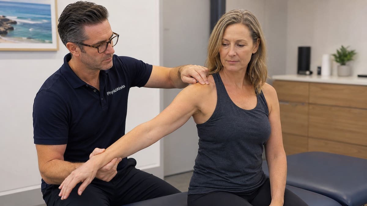
How Is It Checked?
Your physio will check shoulder range, strength, pain pattern, and shoulder blade control. They will also check whether the pain looks more like cuff pain, bursa pain, stiffness, or referred pain.
An X-ray often shows the calcium deposit. Ultrasound can also help. It can check the tendon and bursa. It may also guide a procedure if your doctor suggests one. For a plain-English guide, see MedlinePlus rotator cuff information.
Rotator Cuff Calcific Tendinopathy Treatment
Care depends on pain level, stiffness, work needs, sport goals, and scan findings. Most plans start with simple steps.
| Goal | Plan |
|---|---|
| Ease pain | Change sleep, work, and lift loads for a short time. |
| Restore range | Use gentle drills so the shoulder does not stiffen. |
| Build strength | Progress rotator cuff exercises and shoulder blade control. |
| Return to load | Step back into work, gym, swim, throw, or overhead tasks. |
When Might Other Care Help?
If pain stays high, your care team may discuss other options. These may include shockwave care, image-guided lavage, or an injection. Your GP or sports doctor can help decide whether this fits your case.
Rehab still matters. It helps keep range. It also rebuilds the load you need for daily life.
Should You Keep Exercising?
Usually yes, but lower the load. Avoid sharp pain and heavy overhead work during a flare.
Keep easy motion where you can. Then build lift and sport tasks in steps.
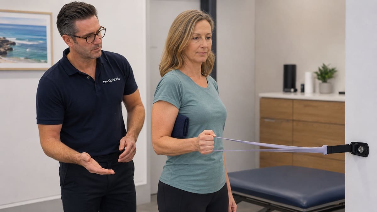
How Physio May Help
Physio aims to match your drills and load to your pain stage. This helps you avoid doing too much too soon. It also helps you avoid losing range.
- Pain control: sleep, lift, work, and gym changes.
- Motion: gentle range drills to reduce guarding.
- Strength: cuff and shoulder blade work.
- Planning: staged return to gym, work, swim, or throw tasks.
Other Shoulder Pages
These pages may help if your pain overlaps with other shoulder problems.
- Rotator cuff tear
- Rotator cuff injury
- Rotator cuff tendinopathy
- Shoulder impingement
- Shoulder bursitis
- Frozen shoulder
- How can you make your rotator cuff heal faster?
Shoulder FAQs
What is this shoulder problem?
It is a shoulder problem where a calcium deposit forms inside a rotator cuff tendon. The supraspinatus tendon is often involved. Pain may flare when the deposit becomes sore or starts to break down.
How long can it last?
Time frames vary. Some people improve over weeks. Others have pain for months, mainly if stiffness builds. A staged plan can help protect range and rebuild strength.
Does it need surgery?
Most people start with non-surgical care. Surgery is usually only discussed when pain and function stay poor after other care has had enough time.
Can shockwave care help?
Shockwave care may suit some people with ongoing pain. It is not the first step for every shoulder. Suitability depends on pain level, scans, goals, and medical advice.
What drills are safe?
Safe drills depend on your pain stage. Early care may use gentle range and light control. Later care often builds cuff strength, shoulder blade control, and load trust.
When should I get checked?
Book an assessment if pain affects sleep, reach, lift, work, or sport. Seek help sooner if pain is severe, range drops fast, or symptoms do not improve in one to two weeks.
What To Do Next
If shoulder pain is affecting sleep, work, or training, book a physio assessment. Your physio can check the likely cause. They can guide safe drills. They can also liaise with your GP if scans or other care may help.
Bring any X-ray, ultrasound, or MRI results to your visit. This helps your plan match your pain stage and goals.
Book your appointment – 24/7
Choose your preferred PhysioWorks clinic and book online.
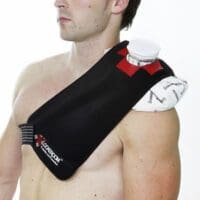
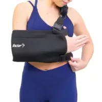
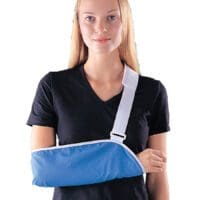
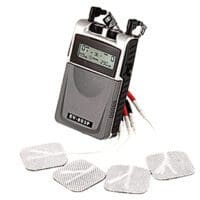
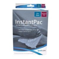
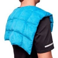
Shoulder Products
These shoulder products are commonly used by our physiotherapists to improve strength, posture, movement, plus assist home exercise programs.
-
-
-
-
-
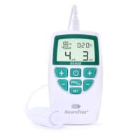
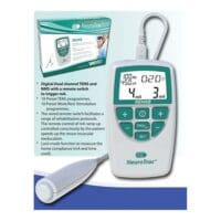
- EMS Machines, Pain Management, TENS Machine, TENS Machine & EMS
NeuroTrac Rehab (TENS Machine + EMS)
$299.95Add to cartQuick View -
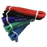
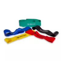
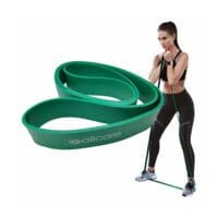 Exercise Equipment, Resistance Band
AllCare Powerloop Bands
$22.00 – $65.00Select optionsQuick View -
-
-
-
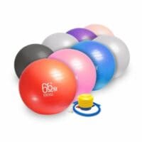
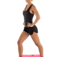
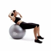 Exercise Balls, Back, Exercise Equipment, Knee, Shoulder
66fit Exercise Balls | Core & Rehab Stability Ball
$32.00 – $57.00Select optionsQuick View -
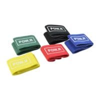 Exercise Equipment, Knee, Resistance Band
POW.R Fabric Mini Loop Bands
$12.00 – $55.00Select optionsQuick View -
-
- Liniments & Gels, Massage Liniments, Pain Management
Fisiocrem
$13.98 – $35.50Select optionsQuick View
Follow PhysioWorks
Get free physiotherapy tips, exercise videos, recovery advice, and blog updates.
| | | | B | | |
References
- Desmeules F, Roy JS, Lafrance S, et al. Rotator Cuff Tendinopathy Diagnosis, Nonsurgical Medical Care, and Rehabilitation: A Clinical Practice Guideline. J Orthop Sports Phys Ther. 2025;55(4):235-274. doi:10.2519/jospt.2025.13182
- Yao Y, Yang G, Jiang S, et al. Treatments for rotator cuff calcific tendinitis: a systematic review and network meta-analysis of randomized-controlled trials. EFORT Open Rev. 2025;10(7):520-533. doi:10.1530/EOR-2024-0078
- Sconza C, Palloni V, Orfei CP, et al. Ultrasound-guided percutaneous lavage for the treatment of rotator cuff calcific tendinopathy: a systematic review with meta-analysis of randomized controlled trials. Eur J Phys Rehabil Med. 2024;60(6):995-1006. doi:10.23736/S1973-9087.24.08513-8
- Catapano M, Robinson DM, Schowalter S, McInnis KC. Clinical evaluation and management of calcific tendinopathy: an evidence-based review. J Osteopath Med. 2022;122(3):141-151. doi:10.1515/jom-2021-0213